Scattered Hyperreflectivity on OCT

Sanjay Sharma MD, MSc (Epid), MBA
Professor of Ophthalmology, Queen’s University
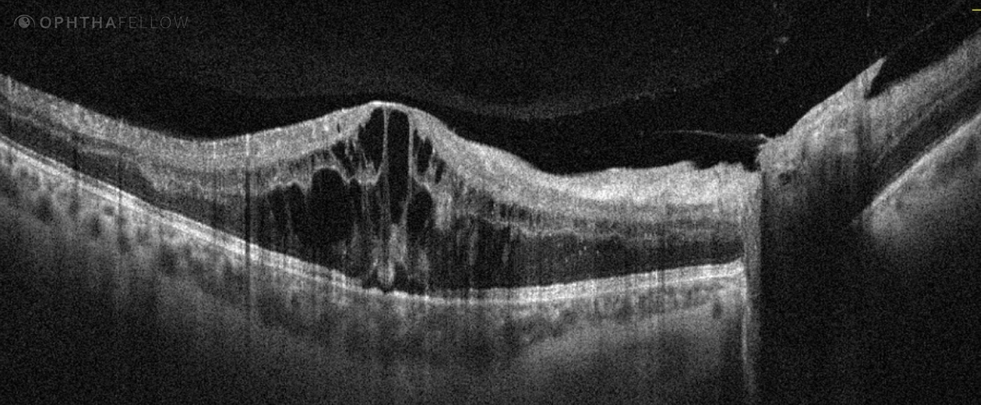
A 30-year-old presents with blurred vision. This image is consistent with which diagnosis?
Analysis
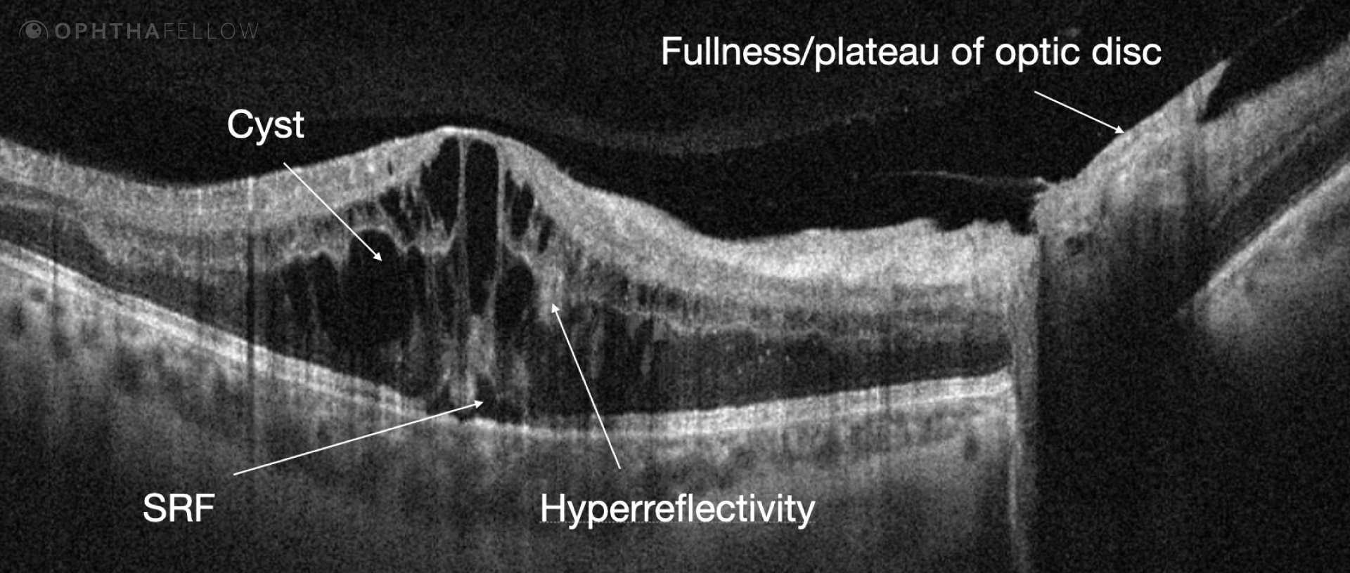
There is a significant dome-shaped elevation in the macular area. There is cystic material in the inner and outer nuclear areas, in addition to significant hyper reflectivity scattered throughout the image. There is also the presence of sub retinal fluid. The optic nerve does not have its typical concavity; this area corresponded to disc neovascularization (see video). Significant schema was seen on OCTA (see video).
The patient was diagnosed with a diabetic macular edema and proliferative retinopathy. They were treated with anti-vegf agents.
Video Analysis
In this tip’s accompanying 15 min. video, we will review:
Clinical Tip
In any patient with DME always look at the nerve on OCT for clues of proliferative retinopathy.